Taking an overview and learning the basic bones of the skeleton will create a solid foundation before looking at each bone and joint in more detail.
Skull
The skull consists of the bones of the head.
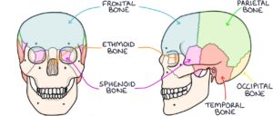
The cranial bones surround the brain. At the front is the frontal bone, at the top is the parietal bone and at the back is the occipital bone. Beside the ear is the temporal bone. Anterior to the temporal bone is the sphenoid bone. The final cranial bone, which is a bit more difficult to visualise, is the ethmoid bone, which is closer to the midline, posterior to the nose and inferior to the frontal bone.
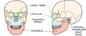
The facial bones form the structure of the face. The bone that forms the bridge of the nose is the nasal bone. The maxilla is the bone that connects the nose, cheekbones and upper teeth. On either side, forming the cheekbones are the zygomatic bones. Finally, the jaw bone is called the mandible. The mandible connects to the temporal bone at the temporomandibular joint (TMJ).
Spine
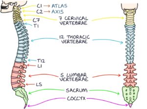
The spine is made up of:
- 7 cervical vertebrae
- 12 thoracic vertebrae
- 5 lumbar vertebrae
- The sacrum
- The coccyx
Vertebrae are numbered from the top down. C1 connects to the base of the skull, followed by C2, C3, C4, C5, C6 and then C7, which connects to the first thoracic vertebra, T1. You then get T1 to T12, L1 to L5, and the sacrum.
C1 and C2 have special names. C1 is called the atlas, and C2 is called the axis.
Upper Limb
The clavicle lies horizontally, between the sternum and the shoulder at the front. It is commonly called the collarbone.
The scapula is the flat, triangular-shaped bone at the back, commonly called the shoulder blade.
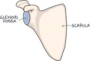
The humerus is the name for the bone of the upper arm. On the scapula, there is a concave area called the glenoid fossa. The head of the humerus meets the glenoid fossa to form the shoulder’s glenohumeral joint.
The radius and ulna bones connect to the humerus at the elbow joint.
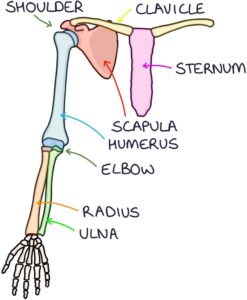
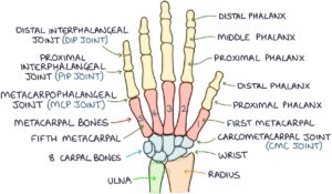
At the wrist, the radius and ulnar connect to the carpal bones. There are 8 carpal bones.
The carpal bones connect to the metacarpal bones. The metacarpals are numbered 1-5, from the thumb to the little finger, meaning the first metacarpal is at the base of the thumb, and the fifth metacarpal is at the base of the little finger.
The fingers and thumb contain the phalanges. Each finger has a proximal phalanx, middle phalanx and distal phalanx. The thumb only has a proximal phalanx and distal phalanx. Moving from the base to the tip of each finger, there is the metacarpophalangeal joint (MCPs), proximal interphalangeal joint (PIP) and distal interphalangeal joint (DIP). At the base of the thumb is the carpometacarpal joint (CMC).
Thorax
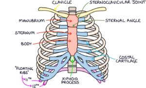
At the top of the thorax is the clavicle. This attaches to the sternum at the sternoclavicular joint. The top part of the sternum is called the manubrium. This attaches to the body of the sternum at the sternal angle. A small bone called the xiphoid process is at the end of the sternum.
There are 12 ribs, one for each thoracic vertebrae. The ribs are labelled 1 to 12, corresponding to the vertebrae they attach to. The costal cartilages connect the ribs to the sternum. The 11th and 12th ribs do not connect to the costal cartilage or the sternum. They are called floating ribs.
Pelvis
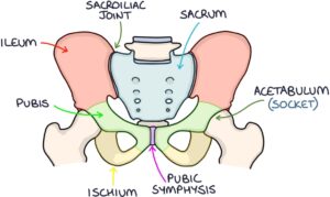
The pelvis comprises three main bones: the ilium, the ischium and the pubis.
At the base of the spine is the sacrum. This attaches to the ilium at the sacroiliac joint. On either side at the front of the pelvis is the pubis bone. The pubis bones join in the centre at the pubic symphysis. Inferiorly, there is the ischium.
The hip joint socket is called the acetabulum and is located at the point where all three pelvis bones meet.
Lower Limb
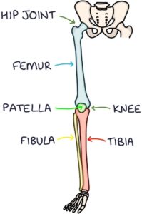
The longest bone in the body is the femur or thigh bone. The head of the femur connects with the acetabulum of the pelvis to form the hip joint.
The femur joins the tibia and fibula of the lower leg to form the knee joint. The tibia is medial, closer to the midline, and the fibula is lateral, on the outer aspect. The patella bone is at the front of the knee joint, commonly called the kneecap.
Ankle and Foot
At the ankle joint, the tibia and fibula meet the tarsal bones of the foot.
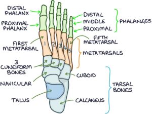
There are 7 tarsal bones:
- Talus (which is the bone that joins with the tibia and fibula at the ankle joint)
- Calcaneus
- Cuboid
- Navicular
- Three cuneiform bones
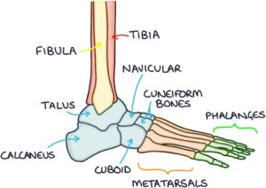
Distal to the tarsal bones are the metatarsals. These are numbered 1-5, with the first metatarsal joining the big toe and the fifth metatarsal joining the little toe.
Distal to the metatarsals are the phalanges. There are proximal, middle and distal phalanges, except for the big toe, which only has a proximal and distal phalanx.
Last updated July 2022
Now, head over to members.zerotofinals.com and test your knowledge of this content. Testing yourself helps identify what you missed and strengthens your understanding and retention.

