
Perthes disease (also called Legg-Calvé-Perthes disease) involves avascular necrosis of the femoral head due to disrupted blood flow. It specifically affects the capital femoral epiphysis, which is the ball that forms the femoral head above the growth plate (physis).
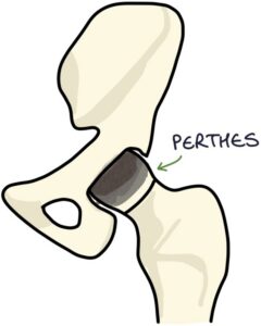
It typically occurs in children aged 4-10 years. It is more common in boys. The severity varies between patients.
It is described as idiopathic, meaning there is no clear cause or trigger. The reason it occurs is unknown.
Over time, revascularisation or neovascularisation of the femoral head occurs, with bone remodelling as it heals. This process often takes 2-3 years.
The main long-term complication is a deformed femoral head that does not fit well in the acetabulum, leading to early hip osteoarthritis. A minority of patients require total hip replacement in adulthood.
Presentation
Perthes disease presents with a gradual onset of:
- Pain in the hip or groin
- Limp
- Restricted hip movements
- Pain may be referred to the knee
There is usually no significant history of trauma.
Investigations
X-ray is the initial investigation for Perthes disease. However, x-rays can be normal, particularly early on.
MRI may be helpful where x-rays are normal, and Perthes disease is still suspected.
Management
Patients should be referred urgently to orthopaedics for management. The key principles of management are to maintain range of motion in the hip and contain the femoral head within the acetabulum while revascularisation and remodelling occur.
Conservative management may involve:
- Activity modification (avoiding high-impact activities)
- Analgesia
- Physiotherapy (to maintain the range of motion)
- Crutches or reduced weight bearing if advised by orthopaedics
Surgery may be used in older children or in severe or complex cases.
Last updated July 2026
Now, head over to members.zerotofinals.com and test your knowledge of this content. Testing yourself helps identify what you missed and strengthens your understanding and retention.
