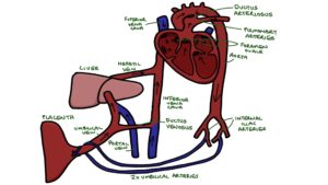
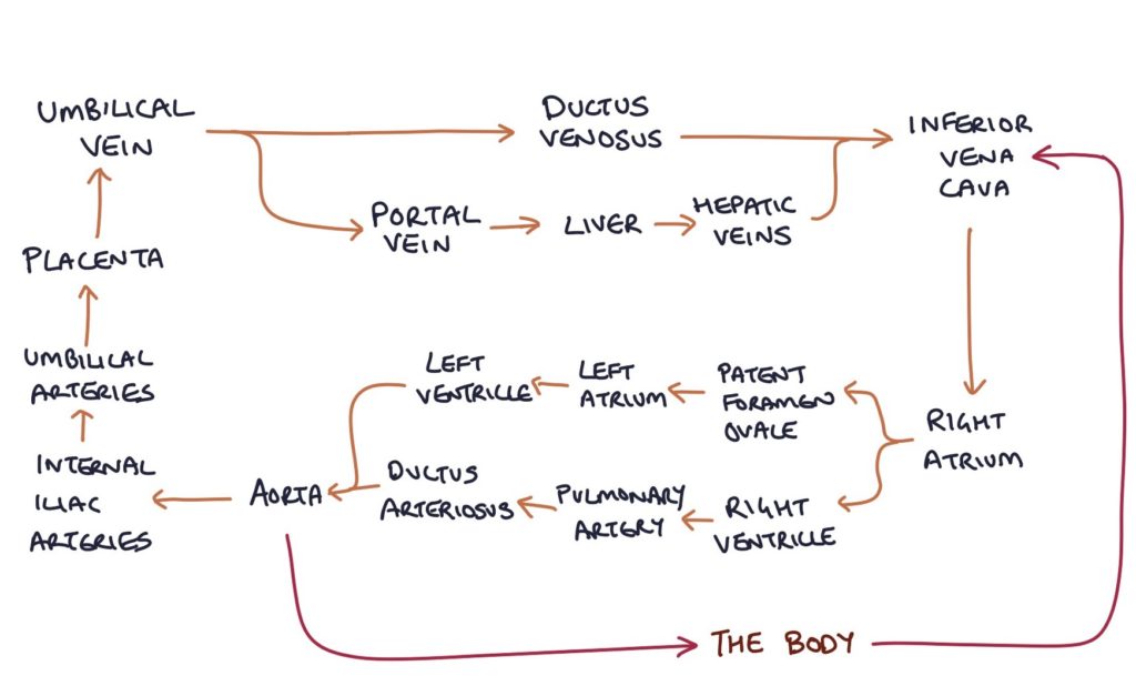
In children and adults, gas exchange happens in the lungs. In the fetus, the lungs are not functional. The placenta is where fetal blood gets oxygenated and waste carbon dioxide is removed.
Fetal blood travels to the placenta via the two umbilical arteries. The umbilical arteries originate from the internal iliac arteries.
Fetal blood travels back from the placenta to the fetus via the umbilical vein.
Fetal Shunts
There are three fetal shunts:
- Ductus venosus
- Foramen ovale
- Ductus arteriosus
The ductus venosus connects the umbilical vein to the inferior vena cava, allowing blood to bypass the liver.
The foramen ovale connects the right atrium with the left atrium, allowing blood to bypass the right ventricle and lungs.
The ductus arteriosus connects the pulmonary artery with the aorta, allowing blood to bypass the lungs.


At Birth
The first breaths after birth expand the alveoli, decreasing the pulmonary vascular resistance. The decrease in pulmonary vascular resistance causes a fall in pressure in the right ventricle and atrium. At this point, the left atrial pressure is greater than the right atrial pressure, which squashes the atrial septum, causing functional closure of the foramen ovale. The foramen ovale is sealed shut within a few weeks, becoming the fossa ovalis.
Prostaglandins are required to keep the ductus arteriosus open. Increased blood oxygenation causes a drop in circulating prostaglandins, causing the closure of the ductus arteriosus, which becomes the ligamentum arteriosum.
When blood stops circulating through the umbilical vein, the ductus venosus stops functioning. The ductus venosus closes structurally a few days later and becomes the ligamentum venosum.
Last updated November 2024
Now, head over to members.zerotofinals.com and test your knowledge of this content. Testing yourself helps identify what you missed and strengthens your understanding and retention.

