Rheumatoid arthritis is an autoimmune condition that causes chronic inflammation in the synovial lining of the joints, tendon sheaths and bursa. It is a type of inflammatory arthritis. Synovial inflammation is called synovitis. Inflammation of the tendons increases the risk of tendon rupture.
Rheumatoid arthritis tends to affect multiple small joints symmetrically across both sides of the body. This pattern is described as symmetrical polyarthritis.
Rheumatoid arthritis is 2-3 times more common in women than men. It most often develops in middle age but can present at any age. Smoking and obesity are risk factors.
A family history increases the risk of rheumatoid arthritis (although there is no clear inheritance pattern). The most common gene associated with rheumatoid arthritis is HLA DR4.
The disease course varies between patients, from mild and remitting to severe and progressive. Disease activity, positive antibodies and erosions on an x-ray predict worse disease.
Antibodies
Rheumatoid factor (RF) is an autoantibody present in around 70% of RA patients. It targets the Fc portion of the immunoglobulin G (IgG). All antibodies (immunoglobulins) have an Fc portion that interacts with other parts of the immune system. Rheumatoid factor causes immune system activation against the patient’s own IgG, resulting in systemic inflammation. Rheumatoid factor is most often IgM but can be other types of immunoglobulin.
Anti-cyclic citrullinated peptide antibodies (anti-CCP antibodies) are more sensitive and specific than rheumatoid factor. They are positive in around 80% of patients with rheumatoid arthritis. They often pre-date the development of rheumatoid arthritis and indicate that a patient will develop the condition at some point.
Presentation
The speed of onset can vary from rapid (e.g., overnight) to gradual (e.g., over months). The three joint symptoms are:
- Pain
- Stiffness
- Swelling
Rheumatoid arthritis typically causes symmetrical distal polyarthritis affecting the small joints of the hands and feet. The most commonly affected joints are:
- Metacarpophalangeal (MCP) joints
- Proximal interphalangeal (PIP) joints
- Wrist
- Metatarsophalangeal (MTP) joints (in the foot)
On palpation of the joints, there will be tenderness and synovial thickening, giving them a “boggy” feeling.
TOM TIP: Rheumatoid arthritis very rarely affects the distal interphalangeal joints. Enlarged and painful distal interphalangeal joints are more likely to represent Heberden’s nodes due to osteoarthritis.
Large joints such as the ankle, knee, hips, and shoulders can also be affected. It can affect the cervical spine (but not the lumbar spine).
Associated systemic symptoms include:
- Fatigue
- Weight loss
- Flu-like illness
- Muscles aches and weakness
TOM TIP: Inflammatory arthritis symptoms are worse with rest and improve with activity. They are worst in the morning. Symptoms of mechanical problems (e.g., osteoarthritis) are worse with activity and improve with rest.
Palindromic Rheumatism
Palindromic rheumatism involves self-limiting episodes of inflammatory arthritis, with pain, stiffness and swelling typically affecting only a few joints. The symptoms last days, then completely resolve. Joints appear normal between episodes. Rheumatoid factor or anti-CCP antibodies may indicate that it will progress to rheumatoid arthritis.
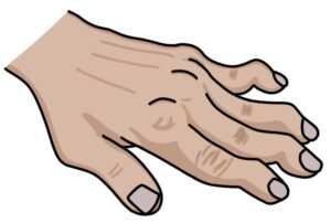
Hand Signs in Advanced Disease
In patients with advanced disease, the hand signs include:
- Z-shaped deformity to the thumb
- Swan neck deformity (hyperextended PIP and flexed DIP)
- Boutonniere deformity (hyperextended DIP and flexed PIP)
- Ulnar deviation of the fingers at the MCP joints
Boutonniere deformity is caused by a tear in the central slip of the extensor components at the proximal interphalangeal (PIP) joint. The central slip connects to the middle phalanx at the PIP, and the lateral bands go around the PIP and connect to the distal phalanx. When the patient tries to straighten their finger, the lateral bands pull on the distal phalanx, causing the distal interphalangeal (DIP) joint to hyperextend and the PIP joint to flex.
Swan neck deformity is the opposite of Boutonnieres deformity. It is caused by an extensor mechanism imbalance, causing flexion of the DIP joint and extension of the PIP joint.
TOM TIP: Effective treatments means it is unusual for rheumatoid arthritis to get to this stage. However, it is worth becoming familiar with these examination findings as they make great patients for OSCEs.
Atlantoaxial Subluxation
Atlantoaxial subluxation occurs in the cervical spine. Synovitis and damage to the ligaments around the odontoid peg of the axis (C2) allow it to shift within the atlas (C1). Subluxation can cause spinal cord compression and is an emergency. This needs to be considered when a patient is having a general anaesthetic and requires intubation. MRI can be used to visualise changes in these areas as part of a pre-operative assessment.
Extra-articular Manifestations
There are many extra-articular manifestations of rheumatoid arthritis:
- Pulmonary fibrosis
- Felty’s syndrome (a triad of rheumatoid arthritis, neutropenia and splenomegaly)
- Sjögren’s syndrome (with dry eyes and dry mouth)
- Anaemia of chronic disease
- Cardiovascular disease
- Eye manifestations
- Rheumatoid nodules (firm, painless lumps under the skin, typically on the elbows and fingers)
- Lymphadenopathy
- Carpel tunnel syndrome
- Amyloidosis
- Bronchiolitis obliterans (small airway destruction and airflow obstruction in the lungs)
- Caplan syndrome (pulmonary nodules in patients with rheumatoid arthritis exposed to coal, silica or asbestos dust)
Eye manifestations related to rheumatoid arthritis and its treatment include:
- Dry eye syndrome (keratoconjunctivitis sicca)
- Episcleritis
- Scleritis
- Keratitis
- Cataracts (secondary to steroids)
- Retinopathy (secondary to hydroxychloroquine)
Diagnosis
The NICE clinical knowledge summaries (updated 2020) recommend an urgent rheumatology referral for patients with persistent synovitis (to be seen within three weeks). They suggest considering an NSAID and arranging baseline bloods while waiting for the specialist assessment.
The investigations that help in the initial assessment are:
- Rheumatoid factor
- Anti-CCP antibodies
- Inflammatory markers, such as C-reactive protein (CRP) and erythrocyte sedimentation rate (ESR)
- X-rays of the hands and feet for bone changes
- Ultrasound or MRI can be used to detect synovitis (useful when clinical findings are unclear)
The diagnosis is based on clinical findings and blood results. The American College of Rheumatology/European League Against Rheumatism (ACR/EULAR) classification criteria from 2010 can be used to make the diagnosis.
X-ray Changes
- Periarticular osteopenia
- Boney erosions
- Soft tissue swelling
- Joint destruction and deformity (in more advanced disease)
Scoring Systems
The Health Assessment Questionnaire (HAQ) measures functional ability. The NICE guidelines (updated 2020) recommend a baseline HAQ score at diagnosis to assess the response to treatment.
The Disease Activity Score 28 Joints (DAS28) score is used in monitoring disease activity and response to treatment. It involves assessing 28 joints and assigning points for:
- Swollen joints
- Tender joints
- The ESR or CRP result
Management
Treatment involves the multidisciplinary team, including rheumatologists, specialist nurses, GPs, physiotherapists, occupational therapists, psychologists and podiatrists.
Starting treatment early improves outcomes. The aim is to induce remission or get as close to remission as possible. C-reactive protein and DAS28 are used to monitor the success of treatment.
Short-term steroids (oral or intramuscular) may be used at initial presentation, when initiating a new treatment and during flares to settle the inflammation and control symptoms quickly.
Treatment is with conventional disease-modifying anti-rheumatic drugs (cDMARDs) and biologic DMARDs:
- Monotherapy with methotrexate, leflunomide or sulfasalazine
- Combination treatment with multiple cDMARDs
- Biologic therapies (usually alongside methotrexate)
Hydroxychloroquine may be used in mild disease and palindromic rheumatism. It is the “mildest” DMARD.
Other cDMARDs include azathioprine, ciclosporin, cyclophosphamide and mycophenolate.
NSAIDs are helpful for pain relief but have associated risks and side effects.
Pregnancy can improve symptoms, but some pregnant women experience a symptom flare. Hydroxychloroquine and sulfasalazine are considered the safest DMARDs in pregnancy (extra folic acid is required with sulfasalazine). Methotrexate and leflunomide are very harmful in pregnancy (teratogenic).
Biological therapies interact with the immune system to reduce inflammation. They have various targets:
- Tumour necrosis factor (TNF) inhibitors (e.g., adalimumab, infliximab, etanercept, golimumab and certolizumab)
- Anti-CD20 on B cells (e.g., rituximab)
- Anti-interleukin-6 inhibitors (e.g., sarilumab and tocilizumab)
- JAK inhibitors (e.g., upadacitinib, tofacitinib and baricitinib)
- T-cell co-stimulation inhibitors (e.g., abatacept)
Tumour necrosis factor is a cytokine involved in stimulating inflammation. Blocking TNF reduces inflammation.
TOM TIP: The main biologics to remember are adalimumab, infliximab and etanercept (TNF inhibitors), and rituximab (a monoclonal antibody that targets the CD20 proteins on the surface of B cells). They cause immunosuppression, increasing the risk of infection, certain cancers (e.g., skin) and reactivation of latent TB.
Orthopaedic surgery used to be an important aspect of management when joint deformities developed. However, cDMARDs and biologics have dramatically reduced the number of patients getting to this stage.
Medication Notes
Methotrexate interferes with folate metabolism and suppresses the immune system. It is given once a week. Folic acid 5mg is taken once a week (on a different day to the methotrexate). Side effects include:
- Mouth ulcers and mucositis
- Liver toxicity
- Bone marrow suppression and leukopenia (low white blood cells)
- Teratogenic (harmful to pregnancy) and needs to be avoided before conception in both women and men
Leflunomide is an immunosuppressant medication that interferes with the production of pyrimidine. Pyrimidine is an important component of RNA and DNA. Side effects include:
- Mouth ulcers and mucositis
- Increased blood pressure
- Liver toxicity
- Bone marrow suppression and leukopenia (low white blood cells)
- Teratogenic (harmful to pregnancy) and needs to be avoided before conception in both women and men
- Peripheral neuropathy
Sulfasalazine is an immunosuppressive and anti-inflammatory medication. The exact mechanism is not clear. Side effects include:
- Orange urine
- Reversible male infertility (reduced sperm count and quality)
- Bone marrow suppression
Hydroxychloroquine is traditionally an antimalarial medication. It suppresses the immune system by interfering with Toll-like receptors, disrupting antigen presentation and increasing the pH in the lysosomes of immune cells. Side effects include:
- Retinal toxicity (reduced visual acuity (macular toxicity)
- Blue-grey skin pigmentation
- Hair lightening (bleaching)
TOM TIP: The unique side effects worth remembering are:
- Methotrexate: Bone marrow suppression and leukopenia, and highly teratogenic
- Leflunomide: Hypertension and peripheral neuropathy
- Sulfasalazine: Orange urine and male infertility (reduces sperm count)
- Hydroxychloroquine: Retinal toxicity, blue-grey skin pigmentation and hair bleaching
- Anti-TNF medications: Reactivation of tuberculosis
- Rituximab: Night sweats and thrombocytopenia
Last updated August 2023
Now, head over to members.zerotofinals.com and test your knowledge of this content. Testing yourself helps identify what you missed and strengthens your understanding and retention.
![]()