Pseudogout is a crystal arthropathy caused by calcium pyrophosphate crystals collecting in the joints. It is formally known as calcium pyrophosphate deposition disease (CPPD). It may also be called chondrocalcinosis.
Presentation
The presentation varies. Many patients are asymptomatic, and it is picked up incidentally on an x-ray. Others may present with chronic pain and stiffness in multiple joints. It can present acutely with a rapid onset of symptoms.
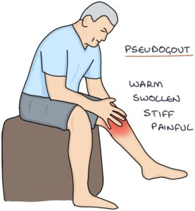
A typical acute presentation of pseudogout is a patient over 65 years old with a rapid-onset hot, swollen, stiff and painful knee. Other commonly affected joints are the shoulders, hips and wrists.
Diagnosis
In any patient presenting with a hot, painful and swollen joint, septic arthritis must be excluded as it is a medical emergency. Symptoms of pseudogout tend to be milder than gout or septic arthritis.
Joint aspiration is used to confirm the diagnosis. Aspirated joint fluid shows calcium pyrophosphate crystals. These are rhomboid-shaped and positively birefringent of polarised light. There should be no bacterial growth.
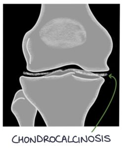
Chondrocalcinosis is the classic x-ray change in pseudogout. The calcium deposits in the joint cartilage show up in a thin white line in the middle of the joint space.
Other joint x-ray changes are similar to osteoarthritis, which can be remembered with the “LOSS” mnemonic:
- L – Loss of joint space
- O – Osteophytes (bone spurs)
- S – Subarticular sclerosis (increased density of the bone along the joint line)
- S – Subchondral cysts (fluid-filled holes in the bone)
Management
Treatment of pseudogout is targeted at symptoms. There are no proven disease-modifying drugs, prophylactic treatments or dietary modifications. Asymptomatic changes on an x-ray do not require any treatment.
Symptoms usually resolve spontaneously over several weeks. Symptomatic management options include:
- NSAIDs (e.g., naproxen) first-line (co-prescribed with a proton pump inhibitor for gastroprotection)
- Colchicine
- Intra-articular steroid injections (septic arthritis must be excluded first)
- Oral steroids
Last updated September 2023
Now, head over to members.zerotofinals.com and test your knowledge of this content. Testing yourself helps identify what you missed and strengthens your understanding and retention.
![]()