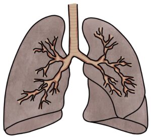
Chronic obstructive pulmonary disease (COPD) involves a long-term, progressive condition involving airway obstruction, chronic bronchitis and emphysema. It is almost always the result of smoking and is largely preventable. While it is not reversible, it is treatable.
Damage to the lung tissues obstructs the flow of air through the airways. Chronic bronchitis refers to long-term symptoms of a cough and sputum production due to inflammation in the bronchi. Emphysema involves damage and dilatation of the alveolar sacs and alveoli, decreasing the surface area for gas exchange.

Unlike asthma, airway obstruction is minimally reversible with bronchodilators, such as salbutamol. Patients are susceptible to exacerbations, during which their lung function worsens. Exacerbations triggered by infection are called infective exacerbations of COPD.
Presentation
A typical presentation of COPD is a long-term smoker with persistent symptoms of:
- Shortness of breath
- Cough
- Sputum production
- Wheeze
- Recurrent respiratory infections, particularly in winter
TOM TIP: COPD does NOT cause clubbing, haemoptysis (coughing up blood) or chest pain. These symptoms should be investigated for a different cause, such as lung cancer, pulmonary fibrosis or heart failure.
MRC Dyspnoea Scale
The MRC (Medical Research Council) Dyspnoea Scale is a 5-point scale for assessing breathlessness:
- Grade 1: Breathless on strenuous exercise
- Grade 2: Breathless on walking uphill
- Grade 3: Breathlessness that slows walking on the flat
- Grade 4: Breathlessness stops them from walking more than 100 meters on the flat
- Grade 5: Unable to leave the house due to breathlessness
Diagnosis
Diagnosis is based on the clinical presentation and spirometry results.
Spirometry will show an obstructive picture with a FEV1:FVC ratio of less than 70%. There is little or no response to reversibility testing with beta-2 agonists (e.g., salbutamol). Reversible obstruction is more suggestive of asthma.
Severity
The severity can be graded using the forced expiratory volume in 1 second (FEV1):
- Stage 1 (mild): FEV1 more than 80% of predicted
- Stage 2 (moderate): FEV1 50-79% of predicted
- Stage 3 (severe): FEV1 30-49% of predicted
- Stage 4 (very severe): FEV1 less than 30% of predicted
Other Investigations
Other investigations include:
- Body mass index at baseline (weight loss occurs in severe disease)
- Chest x-ray to exclude other pathology, such as lung cancer
- Full blood count for polycythaemia (raised haemoglobin due to chronic hypoxia), anaemia and infection
- Sputum culture to assess for chronic infections, such as pseudomonas
- ECG and echocardiogram to assess for heart failure and cor pulmonale
- CT thorax for alternative diagnoses such as fibrosis, cancer or bronchiectasis
- Serum alpha-1 antitrypsin to look for alpha-1 antitrypsin deficiency
- Transfer factor for carbon monoxide (TLCO) tests the diffusion of inhaled gas into the blood (reduced in COPD)
Long-Term Management
Continuing smoking will progressively worsen lung function and prognosis. Smoking cessation services are available.
Patients should have the pneumococcal and annual flu vaccine.
Pulmonary rehabilitation involves a multidisciplinary approach to help improve function and quality of life, including physical training and education.
Initial medical treatment recommended by the NICE guidelines (updated 2019) involves:
- Short-acting beta-2 agonists (e.g., salbutamol)
- Short-acting muscarinic antagonists (e.g., ipratropium bromide)
The second step, when symptoms or exacerbations are still a problem, is determined by whether there are asthmatic or steroid-responsive features, measured by:
- Previous diagnosis of asthma or atopy
- Variation in FEV1 of more than 400mls
- Diurnal variability in peak flow of more than 20%
- Raised blood eosinophil count
Where there are no asthmatic or steroid-responsive features, treatment is a combination of:
- Long-acting beta agonist (LABA)
- Long-acting muscarinic antagonist (LAMA)
Anoro Ellipta, Ultibro Breezhaler and DuaKlir Genuair are examples of LABA and LAMA combination inhalers.
Where there are asthmatic or steroid-responsive features, treatment is a combination of:
- Long-acting beta agonist (LABA)
- Inhaled corticosteroid (ICS)
Fostair, Symbicort and Seretide are examples of LABA and ICS combination inhalers.
The final inhaler step is a combination of a LABA, LAMA and ICS. Trimbow, Trelegy Ellipta and Trixeo Aerosphere are examples of LABA, LAMA and ICS combination inhalers.
In more severe cases, additional options (guided by a specialist) are:
- Nebulisers (e.g., salbutamol or ipratropium)
- Oral theophylline
- Oral mucolytic therapy to break down sputum (e.g., carbocisteine)
- Prophylactic antibiotics (e.g., azithromycin)
- Oral corticosteroids (e.g., prednisolone)
- Oral phosphodiesterase-4 inhibitors (e.g., roflumilast)
- Long-term oxygen therapy at home
- Lung volume reduction surgery (removing damaged lung tissue to improve the function of healthier tissue)
- Palliative care (opiates and other drugs may be used to help breathlessness)
Patients taking azithromycin need ECG and liver function monitoring before and during treatment.
Long-term oxygen therapy (LTOT) is used for severe COPD with chronic hypoxia (sats < 92%), polycythaemia, cyanosis or cor pulmonale. Smoking is a contraindication due to the fire risk.
Cor Pulmonale
Cor pulmonale refers to right-sided heart failure caused by respiratory disease. The increased pressure and resistance in the pulmonary arteries (pulmonary hypertension) limits the right ventricle pumping blood into the pulmonary arteries. This causes back-pressure into the right atrium, vena cava and systemic venous system.
Causes of cor pulmonale are:
- COPD (the most common cause)
- Pulmonary embolism
- Interstitial lung disease
- Cystic fibrosis
- Primary pulmonary hypertension
Often patients with early cor pulmonale are asymptomatic. Symptoms of cor pulmonale include:
- Shortness of breath
- Peripheral oedema
- Breathlessness of exertion
- Syncope (dizziness and fainting)
- Chest pain
Signs of cor pulmonale on examination include:
- Hypoxia
- Cyanosis
- Raised JVP (due to a back-log of blood in the jugular veins)
- Peripheral oedema
- Parasternal heave
- Loud second heart sound
- Murmurs (e.g., pan-systolic in tricuspid regurgitation)
- Hepatomegaly due to back pressure in the hepatic vein (pulsatile in tricuspid regurgitation)
Management of cor pulmonale involves treating the symptoms (e.g., diuretics for oedema) and the underlying cause. Long-term oxygen therapy is often used. The prognosis is poor unless there is a reversible underlying cause.
Acute Exacerbation
An acute COPD exacerbation presents rapidly worsening symptoms, such as cough, shortness of breath, sputum production and wheezing. Viral or bacterial infection often triggers it.
Arterial Blood Gas
An acute exacerbation of COPD typically causes a respiratory acidosis involving:
- Low pH indicates acidosis
- Low pO2 indicates hypoxia and respiratory failure
- Raised pCO2 indicates CO2 retention (hypercapnia)
- Raised bicarbonate indicates chronic retention of CO2
Carbon dioxide (CO2) makes blood acidotic by becoming carbonic acid (H2CO3). Low pH with a raised pCO2 suggests they are acutely retaining CO2, making their blood acidotic, indicating respiratory acidosis.
Raised bicarbonate indicates they chronically retain CO2. Their kidneys have responded by producing more bicarbonate to balance the acidic CO2 and maintain a normal pH. During an acute exacerbation, the kidneys cannot keep up with the rising level of CO2, so the blood becomes acidotic despite a raised bicarbonate.
Other Investigations
Other investigations used during an acute exacerbation include:
- Chest x-ray to look for pneumonia or other pathology
- ECG to look for arrhythmias or evidence of heart strain
- Full blood count to look for infection (raised white blood cells)
- U&E to check electrolytes, which can be affected by infections and medications
- Sputum culture
- Blood cultures in patients with signs of sepsis (e.g., fever)
Oxygen Therapy
Many patients with COPD retain CO2 when treated with oxygen, referred to as oxygen-induced hypercapnia. The mechanism for this is complex and likely involves ventilation-perfusion mismatch and haemoglobin binding less well to CO2 when also bound to oxygen.
Target oxygen saturations of 88-92% are used for patients with COPD at risk of retaining CO2. These may be adjusted to 94-98% when confident they do not retain CO2.
Venturi masks are designed to deliver a specific percentage concentration of oxygen. They allow some of the oxygen to leak out the side of the mask and normal air to be inhaled along with oxygen. Environmental air contains 21% oxygen. Venturi masks deliver 24% (blue), 28% (white), 31% (orange), 35% (yellow), 40% (red) or 60% (green) oxygen.
Management of an Acute Exacerbation
First-line medical treatment of an acute exacerbation of COPD involves:
- Regular inhalers or nebulisers (e.g., salbutamol and ipratropium)
- Steroids (e.g., prednisolone 30 mg once daily for 5 days)
- Antibiotics if there is evidence of infection
Respiratory physiotherapy can be used to help clear sputum.
Additional options in severe cases include:
- IV aminophylline
- Non-invasive ventilation (NIV)
- Intubation and ventilation with admission to intensive care
Doxapram may be used as a respiratory stimulant where NIV or intubation is not appropriate.
Non-Invasive Ventilation
Non-invasive ventilation (NIV) involves using a full face mask, hood (covering the entire head) or a tight-fitting nasal mask to blow air forcefully into the lungs and ventilate them. It is not pleasant for the patient but is much less invasive than intubation and ventilation. It is a valuable middle point between basic oxygen therapy and mechanical ventilation.
NIV involves a cycle of high and low pressure to correspond to the patient’s inspiration and expiration:
- IPAP (inspiratory positive airway pressure) is the pressure during inspiration – where air is forced into the lungs
- EPAP (expiratory positive airway pressure) is the pressure during expiration – stopping the airways from collapsing
NIV is considered when the following inclusion criteria are met:
- Persistent respiratory acidosis (pH < 7.35 and PaCO2 > 6) despite maximal medical treatment
- Potential to recover
- Acceptable to the patient
The decision to initiate it would be made by a registrar or above. The main contraindications are an untreated pneumothorax or any structural abnormality or pathology affecting the face, airway or gastrointestinal tract. Patients should have a chest x-ray before NIV to exclude pneumothorax. A plan should be in place if the NIV fails so that everyone agrees on whether the patient should proceed to intubation, ventilation, and ICU or whether palliative care is more appropriate.
The initial pressures are estimated based on the patient’s body mass. Pressures are measured in cm of water. Potential pressures for an average patient might be:
- IPAP 16-20cm H2O (usually starting at 12 and increasing every 2-5 minutes until the target pressure is reached)
- EPAP 4-6cm H2O
ABGs are monitored closely whilst on NIV (e.g., 1 hour after every change, then 4 hourly until stable). The IPAP is increased by 2-5 cm increments until the acidosis resolves.
Last updated June 2023
Now, head over to members.zerotofinals.com and test your knowledge of this content. Testing yourself helps identify what you missed and strengthens your understanding and retention.

