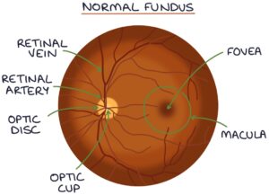
Hypertensive retinopathy involves damage to the small blood vessels in the retina relating to hypertension (high blood pressure). Changes can happen slowly with chronic hypertension or develop quickly in response to malignant hypertension.
Features
Silver wiring or copper wiring is where the walls of the arterioles become thickened and sclerosed and reflect more light on examination.
Arteriovenous nipping (AV nipping) is where the arterioles cause compression of the veins where they cross due to sclerosis and hardening of the arterioles.
Cotton wool spots are caused by ischaemia and infarction in the retina, causing damage to nerve fibres.
Hard exudates are caused by damaged vessels leaking lipids onto the retina.
Retinal haemorrhages are caused by damaged vessels rupturing and releasing blood in the retina. Dot and blot haemorrhages occur deeper, in the inner nuclear layer or outer plexiform layer. Flame haemorrhages occur in the nerve fiber layer.
Papilloedema is caused by ischaemia to the optic nerve, resulting in optic nerve swelling (oedema).

Keith-Wagener Classification
- Stage 1: Mild narrowing of the arterioles
- Stage 2: Focal constriction of blood vessels and AV nicking
- Stage 3: Cotton-wool patches, exudates and haemorrhages
- Stage 4: Papilloedema
Management
Management is focused on controlling blood pressure and managing risk factors (e.g., smoking and blood lipids).
Last updated October 2023
Now, head over to members.zerotofinals.com and test your knowledge of this content. Testing yourself helps identify what you missed and strengthens your understanding and retention.
![]()