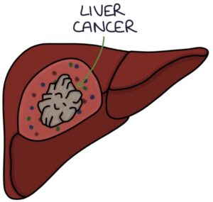
Primary liver cancer is cancer that originates in the liver. The main type of primary liver cancer is hepatocellular carcinoma.
Secondary liver cancer originates outside the liver and metastasises to the liver. Metastasis to the liver can occur with almost any cancer that spreads. It is common to have liver metastases of unknown primary. There is a poor prognosis when there is cancer with liver metastasis.

Risk factors
The main risk factor for hepatocellular carcinoma (HCC) is liver cirrhosis due to:
- Alcohol-related liver disease
- Non-alcoholic fatty liver disease (NAFLD)
- Hepatitis B
- Hepatitis C
- Rarer causes (e.g., primary sclerosing cholangitis)
Patients with liver cirrhosis are offered screening for hepatocellular carcinoma every 6 months with:
- Ultrasound
- Alpha-fetoprotein
Presentation
Liver cancer often remains asymptomatic for a long time, presenting late and making the prognosis poor.
There are non-specific presenting features associated with liver cancer:
- Weight loss
- Abdominal pain
- Anorexia
- Nausea and vomiting
- Jaundice
- Pruritus
- Upper abdominal mass on palpation
Investigations
Relevant investigations in assessing liver cancer are:
- Alpha-fetoprotein (tumour marker for hepatocellular carcinoma)
- Liver ultrasound is the first-line imaging investigation
- CT and MRI scans are used for further assessment and staging of the cancer
- Biopsy is used for histology
Management
Hepatocellular carcinoma has a very poor prognosis unless diagnosed early.
Surgery may be possible in early disease. Resection can be used when the tumour is isolated in a removable liver area. A liver transplant is an option when the tumour is isolated to the liver and the patient meets specific criteria.
Other options for treating liver cancer include:
- Radiofrequency ablation (destroying the tumour cells with heat)
- Microwave ablation (destroying the tumour cells with heat)
- Transarterial chemoembolisation (TACE)
- Radiotherapy
- Targeted drugs (e.g., kinase inhibitors and monoclonal antibodies)
Transarterial chemoembolisation (TACE) is an interventional radiology procedure. A chemotherapy drug is injected into the hepatic artery feeding the tumour, delivering the dose directly to the tumour. This is followed by embolisation of the vessel to block the tumour’s blood supply.
Cholangiocarcinomas
Cholangiocarcinoma is a type of cancer that originates in the bile ducts. The majority are adenocarcinomas. It may affect the bile ducts inside the liver (intrahepatic ducts) or outside the liver (extrahepatic ducts). The most common site is in the perihilar region, where the right and left hepatic ducts have joined to become the common hepatic duct just after leaving the liver.
Cholangiocarcinoma is associated with primary sclerosing cholangitis. However, only 10% of patients with cholangiocarcinoma have primary sclerosing cholangitis. Cholangiocarcinoma usually presents in patients over 50 years old unless related to primary sclerosing cholangitis.
Obstructive jaundice is the key presenting feature to remember. Obstructive jaundice is associated with:
- Pale stools
- Dark urine
- Generalised itching
CA19-9 is a tumour marker for cholangiocarcinoma.
Cholangiocarcinomas have a very poor prognosis unless diagnosed early. Surgical resection is potentially successful in early disease.
TOM TIP: Painless jaundice should make you think of cholangiocarcinoma or cancer of the head of the pancreas. Pancreatic cancer is more common, so this is likely the answer in your exams.
Haemangioma
Haemangiomas are common benign tumours of the liver. They are often found incidentally. They cause no symptoms and have no potential to become cancerous. No treatment or monitoring is required.
Focal Nodular Hyperplasia
Focal nodular hyperplasia is a benign liver tumour made of fibrotic tissue. This is often found incidentally. It is usually asymptomatic and has no malignant potential. It can be related to oestrogen and is more common in women and those on the oral contraceptive pill. No treatment or monitoring is required.
Last updated May 2023
Now, head over to members.zerotofinals.com and test your knowledge of this content. Testing yourself helps identify what you missed and strengthens your understanding and retention.

