Haemochromatosis is an autosomal recessive genetic condition resulting in iron overload. There is excessive total body iron and deposition of iron in tissues. It is an iron storage disorder.
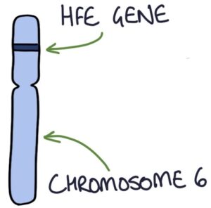
The human haemochromatosis protein (HFE) gene is located on chromosome 6. The majority of cases of haemochromatosis relate to C282Y mutations in this gene. Mutations are required in both copies of the gene (homozygous) since it is an autosomal recessive condition. This gene is important in regulating iron metabolism.

Presentation
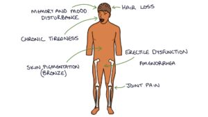
Haemochromatosis usually presents after age 40 when the iron overload becomes symptomatic. It presents later in females due to menstruation acting to eliminate iron from the body regularly. It presents with:
- Chronic tiredness
- Joint pain
- Pigmentation (bronze skin)
- Testicular atrophy
- Erectile dysfunction
- Amenorrhoea (absence of periods in women)
- Cognitive symptoms (memory and mood disturbance)
- Hepatomegaly

Diagnosis
Serum ferritin is the initial investigation. The causes of a raised ferritin are:
- Haemochromatosis
- Infections (it is an acute phase reactant)
- Chronic alcohol consumption
- Non-alcoholic fatty liver disease
- Hepatitis C
- Cancer
Transferrin saturation helps distinguish between high ferritin caused by iron overload (transferrin saturation is high) and other causes (transferrin saturation is normal).
Genetic testing for mutations in the HFE gene is used when the serum ferritin and transferrin saturation are both high.
Liver biopsy with Perl’s stain can be used to establish the iron concentration in the liver. Genetic testing means that a liver biopsy is not usually necessary for establishing a diagnosis. It may help stage the fibrosis and exclude other liver pathology.
MRI can give a detailed picture and quantify the iron concentration in the liver (avoiding a biopsy).
Complications
Haemochromatosis has a long list of complications:
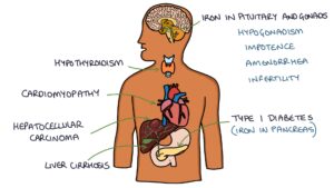
- Secondary diabetes (iron affects the functioning of the pancreas)
- Liver cirrhosis
- Endocrine and sexual problems (hypogonadism, erectile dysfunction, amenorrhea and reduced fertility)
- Cardiomyopathy (iron deposits in the heart)
- Hepatocellular carcinoma
- Hypothyroidism (iron deposits in the thyroid)
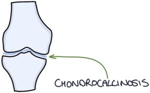
- Chondrocalcinosis (calcium pyrophosphate deposits in joints) causes arthritis

Management
Management involves:
- Venesection (regularly removing blood to remove excess iron – initially weekly)
- Monitoring serum ferritin
- Monitoring and treating complications
Last updated May 2023
Now, head over to members.zerotofinals.com and test your knowledge of this content. Testing yourself helps identify what you missed and strengthens your understanding and retention.


