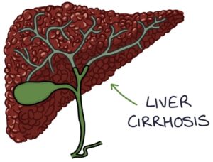
Liver cirrhosis is the result of chronic inflammation and damage to liver cells. The functional liver cells are replaced with scar tissue (fibrosis). Nodules of scar tissue form within the liver.
Fibrosis affects the structure and blood flow through the liver, increasing the resistance in the vessels leading into the liver. This increased resistance and pressure in the portal system is called portal hypertension.
Causes
The four most common causes of liver cirrhosis are:
- Alcohol-related liver disease
- Non-alcoholic fatty liver disease (NAFLD)
- Hepatitis B
- Hepatitis C
Cirrhosis also has many rarer causes:
- Autoimmune hepatitis
- Primary biliary cirrhosis
- Haemochromatosis
- Wilsons disease
- Alpha-1 antitrypsin deficiency
- Cystic fibrosis
- Drugs (e.g., amiodarone, methotrexate and sodium valproate)
Examination Findings
Signs of liver cirrhosis on examination include:
- Cachexia (wasting of the body and muscles)
- Jaundice caused by raised bilirubin
- Hepatomegaly (enlargement of the liver)
- Small nodular liver as it becomes more cirrhotic
- Splenomegaly due to portal hypertension
- Spider naevi (telangiectasia with a central arteriole and small vessels radiating away)
- Palmar erythema caused by elevated oestrogen levels
- Gynaecomastia and testicular atrophy in males due to endocrine dysfunction
- Bruising due to abnormal clotting
- Excoriations (scratches on the skin due to itching)
- Ascites (fluid in the peritoneal cavity)
- Caput medusae (distended paraumbilical veins due to portal hypertension)
- Leukonychia (white fingernails) associated with hypoalbuminaemia
- Asterixis (“flapping tremor”) in decompensated liver disease
Non-Invasive Liver Screen
Abnormal liver function tests without a clear cause require a non-invasive liver screen, which includes:
- Ultrasound liver (used to diagnose fatty liver)
- Hepatitis B and C serology
- Autoantibodies (autoimmune hepatitis, primary biliary cirrhosis and primary sclerosing cholangitis)
- Immunoglobulins (autoimmune hepatitis and primary biliary cirrhosis)
- Caeruloplasmin (Wilsons disease)
- Alpha-1 antitrypsin levels (alpha-1 antitrypsin deficiency)
- Ferritin and transferrin saturation (hereditary haemochromatosis)
Autoantibodies relevant to liver disease include:
- Antinuclear antibodies (ANA)
- Smooth muscle antibodies (SMA)
- Antimitochondrial antibodies (AMA)
- Antibodies to liver kidney microsome type-1 (LKM-1)
Blood Tests
Liver function tests (LFTs) may be normal in cirrhosis. However, in decompensated cirrhosis, all the liver markers become deranged, with raised:
- Bilirubin
- Alanine transaminase (ALT)
- Aspartate transferase (AST)
- Alkaline phosphatase (ALP)
Other blood results include:
- Low albumin due to reduced synthetic function of the liver
- Increased prothrombin time due to reduced synthetic function of the liver (reduced production of clotting factors)
- Thrombocytopenia (low platelets) is a common finding and indicates more advanced disease
- Hyponatraemia (low sodium) occurs with fluid retention in severe liver disease
- Urea and creatinine become deranged in hepatorenal syndrome
- Alpha-fetoprotein is a tumour marker for hepatocellular carcinoma
The enhanced liver fibrosis (ELF) blood test is the first-line investigation for assessing fibrosis in non-alcoholic fatty liver disease. It is not used in patients with other causes of liver disease. It measures three markers (HA, PIIINP and TIMP-1) and uses an algorithm to provide a result that indicates whether they have advanced fibrosis of the liver:
- 10.51 or above – advanced fibrosis
- Under 10.51 – unlikely advanced fibrosis (NICE recommend rechecking every 3 years in NAFLD)
Ultrasound
Ultrasound is used to diagnose non-alcoholic fatty liver disease (once other causes are excluded). Fatty changes appear as increased echogenicity.
In liver cirrhosis, an ultrasound may show:
- Nodularity of the surface of the liver
- A “corkscrew” appearance to the hepatic arteries with increased flow as they compensate for reduced portal flow
- Enlarged portal vein with reduced flow
- Ascites
- Splenomegaly
Ultrasound is used as a screening tool for hepatocellular carcinoma (alongside alpha-fetoprotein).
Transient Elastography
Transient elastography (“FibroScan”) can be used to assess the stiffness of the liver using high-frequency sound waves. It helps determine the degree of fibrosis (scarring) to test for liver cirrhosis. It is used in patients at risk of cirrhosis:
- Alcohol-related liver disease
- Heavy alcohol drinkers (men drinking more than 50 units or women drinking more than 35 units per week)
- Non-alcoholic fatty liver disease and advanced liver fibrosis (score 10.51 or more on the ELF blood test)
- Hepatitis C
- Chronic hepatitis B
Other Investigations
Endoscopy can be used to assess for and treat oesophageal varices when portal hypertension is suspected.
CT and MRI can be used to look for hepatocellular carcinoma, hepatosplenomegaly, abnormal blood vessel changes and ascites.
Liver biopsy can be used to confirm the diagnosis of cirrhosis.
MELD Score
NICE recommend using the MELD (Model for End-Stage Liver Disease) score every 6 months in patients with compensated cirrhosis. The formula considers the bilirubin, creatinine, INR and sodium and whether they require dialysis, giving an estimated 3-month mortality as a percentage.
Child-Pugh Score
The Child-Pugh scores uses 5 factors to assess the severity of cirrhosis and the prognosis. Each factor is considered and scored 1, 2 or 3. The minimum overall score is 5 (scoring 1 for each factor), and the maximum is 15 (scoring 3 for each factor). You can remember the features with the “ABCDE” mnemonic:
- A – Albumin
- B – Bilirubin
- C – Clotting (INR)
- D – Dilation (ascites)
- E – Encephalopathy
General Management
There are four principles of management:
- Treating the underlying cause
- Monitoring for complications
- Managing complications
- Liver transplant
The underlying cause needs to be addressed. For example:
- Stop drinking alcohol
- Lifestyle changes for non-alcohol fatty liver disease
- Antiviral drugs for hepatitis C
- Immunosuppressants for autoimmune hepatitis
Monitoring for complications involves:
- MELD score every 6 months
- Ultrasound and alpha-fetoprotein every 6 months for hepatocellular carcinoma
- Endoscopy every 3 years for oesophageal varices
Liver transplantation is generally considered when there are features of decompensated liver disease. The four key features can be remembered with the “AHOY” mnemonic:
- A – Ascites
- H – Hepatic encephalopathy
- O – Oesophageal varices bleeding
- Y – Yellow (jaundice)
Complications and Prognosis
The course of the disease is variable. Overall, 5-year survival is about 50% once cirrhosis has developed. The MELD score or Child-Pugh score can be used as prognostic tools.
There are several important complications of cirrhosis:
- Malnutrition and muscle wasting
- Portal hypertension, oesophageal varices and bleeding varices
- Ascites and spontaneous bacterial peritonitis
- Hepatorenal syndrome
- Hepatic encephalopathy
- Hepatocellular carcinoma
Malnutrition
Cirrhosis leads to malnutrition and muscle wasting. Patients often have a loss of appetite resulting in reduced intake. Cirrhosis affects protein metabolism in the liver and reduces the amount of protein the liver produces. It also disrupts the ability of the liver to store glucose as glycogen and release it when required. Overall, less protein is available for maintaining muscle tissue and muscle tissue is broken down for use as fuel.
Management involves nutritional support guided by a dietician, with:
- Regular meals
- High protein and calorie intake
- Reduced sodium intake to minimise fluid retention
- Avoiding alcohol
Portal Hypertension and Varices
The portal vein comes from the superior mesenteric and splenic veins and delivers blood to the liver. Liver cirrhosis increases the resistance to blood flow in the liver. As a result, there is increased back pressure on the portal system. This is called portal hypertension. The back pressure of blood results in splenomegaly.
Back pressure in the portal system causes swollen and tortuous vessels at sites where collaterals form between the portal and systemic venous systems. These collaterals can occur at several locations, notably the:
- Distal oesophagus (oesophageal varices)
- Anterior abdominal wall (caput medusae)
Varices are asymptomatic until they start bleeding. Due to the high blood flow, bleeding from varices can cause patients to exsanguinate (bleed out) very quickly.
Prophylaxis of bleeding in stable oesophageal varices involves:
- Non-selective beta blockers (e.g., propranolol) first-line
- Variceal band ligation (if beta blockers are contraindicated)
Variceal band ligation involves a rubber band wrapped around the base of the varices, cutting off the blood flow through the vessels.
Bleeding Oesophageal Varices
Bleeding oesophageal varices is a life-threatening emergency. Initial management involves:
- Immediate senior help
- Consider blood transfusion (activate the major haemorrhage protocol)
- Treat any coagulopathy (e.g., with fresh frozen plasma)
- Vasopressin analogues (e.g., terlipressin or somatostatin) cause vasoconstriction and slow bleeding
- Prophylactic broad-spectrum antibiotics (shown to reduce mortality)
- Urgent endoscopy with variceal band ligation
- Consider intubation and intensive care
Other options to control the bleeding include:
- Sengstaken-Blakemore tube (an inflatable tube inserted into the oesophagus to tamponade the bleeding varices)
- Transjugular intrahepatic portosystemic shunt (TIPS)
Transjugular intrahepatic portosystemic shunt (TIPS) is a technique where an interventional radiologist inserts a wire under x-ray guidance into the jugular vein, down the vena cava and into the liver via the hepatic vein. A connection is made through the liver between the hepatic vein and portal vein, and a stent is inserted. This allows blood to flow directly from the portal vein to the hepatic vein, relieving the pressure in the portal system. The two main indications are:
- Bleeding oesophageal varices
- Refractory ascites
Ascites
Ascites refers to fluid in the peritoneal cavity. The increased pressure in the portal system causes fluid to leak out of the capillaries in the liver and other abdominal organs into the peritoneal cavity. The drop in circulating volume caused by fluid loss into the peritoneal cavity causes reduced blood pressure in the kidneys. The kidneys sense this lower pressure and release renin, which leads to increased aldosterone secretion via the renin-angiotensin-aldosterone system. Increased aldosterone causes the reabsorption of fluid and sodium in the kidneys, leading to fluid and sodium retention. Cirrhosis causes transudative (low protein content) ascites.
Management options include:
- Low sodium diet
- Aldosterone antagonists (e.g., spironolactone)
- Paracentesis (ascitic tap or ascitic drain)
- Prophylactic antibiotics (ciprofloxacin or norfloxacin) when there is <15 g/litre of protein in the ascitic fluid
- Transjugular intrahepatic portosystemic shunt (TIPS) is considered in refractory ascites
- Liver transplantation is considered in refractory ascites
Spontaneous Bacterial Peritonitis
Spontaneous bacterial peritonitis (SBP) occurs in 10-20% of patients with ascites. It has a mortality of 10-20%. It involves an infection developing in the ascitic fluid and peritoneal lining without a clear source of infection (e.g., an ascitic drain or bowel perforation).
Spontaneous bacterial peritonitis can be asymptomatic. Presenting features include:
- Fever
- Abdominal pain
- Deranged bloods (raised WBC, CRP, creatinine or metabolic acidosis)
- Ileus (reduced movement in the intestines)
- Hypotension
The most common organisms are:
- Escherichia coli
- Klebsiella pneumoniae
Management involves:
- Taking a sample of ascitic fluid for culture before giving antibiotics
- Intravenous broad-spectrum antibiotics according to local policies (e.g., piperacillin with tazobactam)
Hepatorenal Syndrome
Hepatorenal syndrome involves impaired kidney function caused by changes in the blood flow to the kidneys relating to liver cirrhosis and portal hypertension.
The exact pathophysiology is still being debated. A simplified version is that portal hypertension causes the portal vessels to release vasodilators, which cause significant vasodilation in the splanchnic circulation (the vessels supplying the gastrointestinal organs). Vasodilation leads to reduced blood pressure. The kidneys respond to the reduced pressure by activating the renin-angiotensin-aldosterone system, which leads to vasoconstriction of the renal vessels. Renal vasoconstriction combined with low systemic pressure results in the kidneys being starved of blood and significantly reduced kidney function.
Hepatorenal syndrome has a poor prognosis unless the patient has a liver transplant.
Hepatic Encephalopathy
Hepatic encephalopathy is also known as portosystemic encephalopathy. It is thought to be caused by the build-up of neurotoxic substances that affect the brain.
One toxin particularly worth remembering is ammonia, produced by intestinal bacteria when they break down proteins. Ammonia is absorbed in the intestines. There are two reasons that ammonia builds up in the blood in patients with cirrhosis: Firstly, the liver cells’ functional impairment prevents them from metabolising the ammonia into harmless waste products. Secondly, collateral vessels between the portal and systemic circulation mean that the ammonia bypasses the liver and enters the systemic system directly.
Acutely, hepatic encephalopathy presents with reduced consciousness and confusion. It can present more chronically with changes to personality, memory and mood.
Factors that can trigger or worsen hepatic encephalopathy are:
- Constipation
- Dehydration
- Electrolyte disturbance
- Infection
- Gastrointestinal bleeding
- High protein diet
- Medications (particularly sedative medications)
Management involves:
- Lactulose (aiming for 2-3 soft stools daily)
- Antibiotics (e.g., rifaximin) to reduce the number of intestinal bacteria producing ammonia
- Nutritional support (nasogastric feeding may be required)
Lactulose works in several ways to reduce ammonia:
- Speeds up transit time and reduces constipation (the laxative effect clearing the ammonia before it is absorbed)
- Promotes bacterial uptake of ammonia to be used for protein synthesis
- Changes the pH of the contents of the intestine to become more acidic, killing ammonia-producing bacteria
Rifaximin is the usual choice of antibiotic as it is poorly absorbed and stays in the gastrointestinal tract. Neomycin and metronidazole are alternatives.
Last updated May 2023
Now, head over to members.zerotofinals.com and test your knowledge of this content. Testing yourself helps identify what you missed and strengthens your understanding and retention.
![]()