Sickle cell disease is a genetic condition that causes sickle-shaped red blood cells.
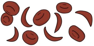
The abnormal shape makes the red blood cells more fragile and easily destroyed, leading to haemolytic anaemia. Patients with sickle cell disease are prone to various sickle cell crises.
Pathophysiology
Haemoglobin is the protein in red blood cells that transports oxygen. During fetal development, at around 32-36 weeks gestation, fetal haemoglobin (HbF) production decreases, and adult haemoglobin (HbA) increases. There is a gradual transition from HbF to HbA. At birth, around 70–80% is HbF and 20–30% is HbA. By 6 months, very little HbF is produced, and red blood cells contain almost entirely HbA.
Patients with sickle-cell disease have an abnormal variant called haemoglobin S (HbS). HbS results in sickle-shaped red blood cells.
It is an autosomal recessive condition caused by a mutation in the beta-globin gene on chromosome 11. One abnormal copy of the gene results in sickle-cell trait. Patients with sickle-cell trait are usually asymptomatic. They are carriers of the condition. Two abnormal copies result in sickle-cell disease.
Relation to Malaria
Sickle cell disease is more common in patients from areas traditionally affected by malaria, such as Africa, India, the Middle East and the Caribbean. Having one copy of the gene (sickle cell trait) reduces the severity of malaria. As a result, patients with sickle cell trait are more likely to survive malaria and pass on their genes. Therefore, there is a selective advantage to having the sickle cell gene in areas of malaria, making it more common.
Screening
Sickle cell disease is tested for on the newborn blood spot screening test at around five days of age.
Pregnant women at high risk of being carriers of the sickle cell gene are offered testing.
Complications
- Anaemia
- Increased risk of infection
- Chronic kidney disease
- Sickle cell crises
- Acute chest syndrome
- Stroke
- Avascular necrosis in large joints such as the hip
- Pulmonary hypertension
- Gallstones
- Priapism (painful and persistent penile erections)
Sickle Cell Crisis
Sickle cell crisis refers to a spectrum of acute exacerbations caused by sickle cell disease. These range from mild to life-threatening. They can occur spontaneously or be triggered by dehydration, infection, stress or cold weather.
There is no specific treatment for sickle cell crisis. They are managed supportively, with:
- Low threshold for admission to hospital
- Treating infections that may have triggered the crisis
- Keeping warm
- Good hydration (IV fluids may be required)
- Analgesia (NSAIDs should be avoided where there is renal impairment)
Vaso-Occlusive Crisis
Vaso-occlusive crisis (VOC), also known as painful crisis, is the most common type of sickle cell crisis. It is caused by sickle-shaped red blood cells clogging capillaries, leading to distal ischaemia.
It typically presents with pain and swelling in the hands or feet, but can affect the chest, back, or other body areas. It can be associated with fever.
It can cause priapism in men by trapping blood in the penis, causing a painful and persistent erection. Priapism is a urological emergency, treated by aspirating blood from the penis.
Splenic Sequestration Crisis
Splenic sequestration crisis is caused by red blood cells trapping blood in the spleen. It causes an acutely enlarged and painful spleen. Blood pooling in the spleen can lead to severe anaemia and hypovolaemic shock.
Splenic sequestration crisis is considered an emergency. Management is supportive, with blood transfusions and fluid resuscitation to treat anaemia and shock.
Recurrent vaso-occlusive episodes in the spleen can lead to splenic infarction, hyposplenism, and infections with encapsulated bacteria (e.g., Streptococcus pneumoniae and Haemophilus influenzae).
Splenectomy prevents sequestration crises and may be used in recurrent cases.
Aplastic Crisis
Aplastic crisis involves a temporary absence of red blood cell production. It may be triggered by parvovirus B19 infection.
It leads to significant anaemia. Management is supportive, with blood transfusions if necessary. It usually resolves spontaneously within around a week.
Acute Chest Syndrome
Acute chest syndrome occurs when the vessels supplying the lungs become clogged with red blood cells. A vaso-occlusive crisis, fat embolism or infection can trigger it.
Acute chest syndrome presents with fever, shortness of breath, chest pain, cough and hypoxia. A chest x-ray will show pulmonary infiltrates.
Acute chest syndrome is a medical emergency with high mortality. It requires prompt supportive management and treatment of the underlying cause, with:
- Analgesia
- Good hydration (IV fluids may be required)
- Antibiotics for infections
- Exchange transfusions
- Incentive spirometry using a machine that encourages effective and deep breathing
- Respiratory support with oxygen, non-invasive ventilation or mechanical ventilation
General Management
A specialist MDT will manage sickle cell disease. The general principles are:
- Avoid triggers for crises, such as dehydration
- Up-to-date vaccinations
- Antibiotic prophylaxis to protect against infection, typically with penicillin V (phenoxymethylpenicillin)
- Hydroxycarbamide (stimulates HbF)
- Bone marrow transplant can be curative
- Crizanlizumab (monoclonal antibody targeting P-selectin) (age 16 and older)
Hydroxycarbamide works by stimulating the production of fetal haemoglobin (HbF). Fetal haemoglobin does not lead to sickling of red blood cells (unlike HbS). It reduces the frequency of vaso-occlusive crises, improves anaemia and may extend lifespan.
Crizanlizumab is a monoclonal antibody that targets P-selectin. P-selectin is an adhesion molecule found on endothelial cells on the inside walls of blood vessels and on platelets. It prevents red blood cells from sticking to the blood vessel wall and reduces the frequency of vaso-occlusive crises.
Last updated March 2026
Now, head over to members.zerotofinals.com and test your knowledge of this content. Testing yourself helps identify what you missed and strengthens your understanding and retention.
![]()