Thyroid function tests can be used to check for abnormal thyroid function and determine the cause.
Thyroid Axis Physiology
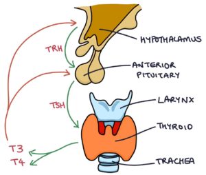
Thyroid hormone levels are controlled by two structures in the brain called the hypothalamus and the pituitary gland, specifically the anterior part of the pituitary.
The hypothalamus releases thyrotropin-releasing hormone (TRH). TRH stimulates the anterior pituitary to release thyroid-stimulating hormone (TSH). TSH stimulates the thyroid gland to release triiodothyronine (T3) and thyroxine (T4).
The hypothalamus and anterior pituitary respond to T3 and T4 by suppressing the release of TRH and TSH, resulting in lower amounts of T3 and T4. The lower T3 and T4 offer less suppression of TRH and TSH, causing more of these hormones to be released, resulting in a rise of T3 and T4. This way, the thyroid hormone level is closely regulated to keep it within normal limits.
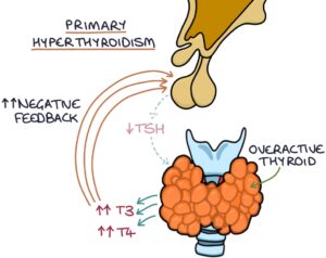
When the end hormone (e.g., T3 and T4) suppresses the release of the controlling hormones (e.g., TRH and TSH), this is called negative feedback.
Hormone Tests
Thyroid-stimulating hormone (TSH) is used as a screening test for thyroid disease. When TSH is abnormal, triiodothyronine (T3) and thyroxine (T4) can be measured to gain more information.
Primary hyperthyroidism is where the thyroid behaves abnormally and produces excessive thyroid hormones. TSH is suppressed by the high T3 and T4, causing a low TSH level. The top causes of primary hyperthyroidism can be remembered with the “GIST” mnemonic:
- G – Graves’ disease
- I – Inflammation (thyroiditis)
- S – Solitary toxic thyroid nodule
- T – Toxic multinodular goitre
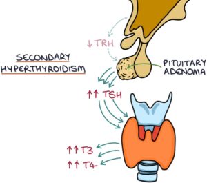
Secondary hyperthyroidism is where the pituitary behaves abnormally and produces excessive TSH (e.g., pituitary adenoma), stimulating the thyroid gland to produce excessive thyroid hormones. TSH, T3 and T4 will all be raised.
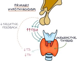
Primary hypothyroidism is where the thyroid behaves abnormally and produces inadequate thyroid hormones. Negative feedback is absent, resulting in increased production of TSH. TSH is raised, and T3 and T4 are low. The top causes of primary hypothyroidism are:
- Hashimoto’s thyroiditis
- Iodine deficiency
- Treatments for hyperthyroidism
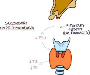
Secondary hypothyroidism is where the pituitary behaves abnormally and produces inadequate TSH (e.g., after surgical removal of the pituitary), resulting in under-stimulation of the thyroid gland and insufficient thyroid hormones. TSH, T3 and T4 will all be low.
|
TSH |
T3 and T4 |
|
|
Primary Hyperthyroidism |
Low |
High |
|
Secondary Hyperthyroidism |
High |
High |
|
Primary Hypothyroidism |
High |
Low |
|
Secondary Hypothyroidism |
Low |
Low |
Antibodies
Anti-thyroid peroxidase (anti-TPO) antibodies are antibodies against the thyroid gland. They are the most relevant thyroid autoantibody in autoimmune thyroid disease. They are usually present in Grave’s disease and Hashimoto’s thyroiditis.
Anti-thyroglobulin (anti-Tg) antibodies are antibodies against thyroglobulin, a protein produced and extensively present in the thyroid gland. They can be present in normal individuals without thyroid pathology. They are usually raised with Grave’s disease, Hashimoto’s thyroiditis and thyroid cancer.
TSH receptor antibodies are autoantibodies that mimic TSH, bind to the TSH receptor and stimulate thyroid hormone release. They cause Grave’s disease and will therefore be present in this condition.
Imaging
Ultrasound of the thyroid gland helps diagnose thyroid nodules and distinguish between cystic (fluid-filled) and solid nodules. Ultrasound can also be used to guide a biopsy of a thyroid lesion.
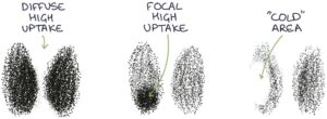
Radioisotope scans are used to investigate hyperthyroidism and thyroid cancers. Radioactive iodine is given orally or intravenously and travels to the thyroid, where it is taken up by the thyroid cells. Iodine used by thyroid cells to produce thyroid hormones. The more active the thyroid cells, the faster the radioactive iodine is taken up. A gamma camera detects gamma rays emitted from the radioactive iodine. The more gamma rays emitted from an area, the more radioactive iodine has been taken up. This gives functional information about the thyroid gland:
- Diffuse high uptake is found in Grave’s Disease
- Focal high uptake is found in toxic multinodular goitre and adenomas
- “Cold” areas (abnormally low uptake) can indicate thyroid cancer
Last updated July 2024
Now, head over to members.zerotofinals.com and test your knowledge of this content. Testing yourself helps identify what you missed and strengthens your understanding and retention.
![]()