Bleeding from the upper gastrointestinal tract is a relatively common medical emergency. It involves bleeding from the oesophagus, stomach or duodenum.
Causes
The key sources of bleeding are:
- Peptic ulcers (the most common cause)
- Mallory-Weiss tear (a tear of the oesophageal mucosa)
- Oesophageal varices (secondary to portal hypertension in liver cirrhosis)
- Stomach cancers
Presentation
The presenting features of an upper gastrointestinal bleed are:
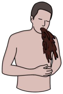
- Haematemesis (vomiting blood)
- Coffee ground vomit (caused by vomiting digested blood with the appearance of coffee grounds)
- Melaena (tar-like, black, greasy and offensive stools caused by digested blood)
Haemodynamic instability occurs with significant blood loss, causing low blood pressure, tachycardia and other signs of shock. Young, fit patients may compensate well with normal observations until they have lost a lot of blood.
Peptic ulcers are associated with a history of epigastric pain and dyspepsia. They may be taking non-steroidal anti-inflammatory drugs (NSAIDs).
Mallory-Weiss tears tend to occur after heavy retching or vomiting, which may be caused by binge drinking, gastroenteritis or hyperemesis gravidarum (in early pregnancy).
Oesophageal varices are associated with liver cirrhosis and portal hypertension. The patient will have signs of these conditions, such as ascites, jaundice and caput medusae.
Stomach cancer is associated with a history of weight loss, epigastric pain, treatment-resistant dyspepsia, low haemoglobin (anaemia) and a raised platelet count.
Glasgow-Blatchford Bleeding Score
The Glasgow-Blatchford score is used at the initial presentation in suspected upper GI bleed. It estimates the risk of the patient having an upper GI bleed. A score above 0 indicates a high risk for an upper GI bleed. The NICE guidelines (updated 2016) suggest considering early discharge in patients with a score of 0.
The easiest way to calculate the score is using an online calculator. It takes into account:
- Haemoglobin (falls in upper GI bleeding)
- Urea (rises in upper GI bleeding)
- Systolic blood pressure
- Heart rate
- Presence of melaena (black, tarry stools)
- Syncope (loss of consciousness)
- Liver disease
- Heart failure
TOM TIP: Acid and digestive enzymes break down blood in the upper GI tract. One of the breakdown products is urea, which is then absorbed in the intestines, causing a rise in blood urea. The association between upper GI bleeding and increased blood urea is a key fact worth remembering.
Rockall Score
The Rockall score is used after endoscopy to estimate the risk of rebleeding and mortality. It takes into account:
- Age
- Features of shock (e.g., tachycardia or hypotension)
- Co-morbidities
- Cause of bleeding (e.g., Mallory-Weiss tear or malignancy)
- Endoscopic findings of recent bleeding (e.g., clots and visible bleeding vessels)
Management
As with any medical emergency, get senior support early and follow the local policies.
The initial management can be remembered with the ABATED mnemonic:
- A – ABCDE approach to immediate resuscitation
- B – Bloods
- A – Access (ideally 2 x large bore cannula)
- T – Transfusions are required
- E – Endoscopy (within 24 hours)
- D – Drugs (stop anticoagulants and NSAIDs)
Send bloods for:
- Haemoglobin (FBC)
- Urea (U&Es)
- Coagulation (INR and FBC for platelets)
- Liver disease (LFTs)
- Crossmatch 2 units of blood
TOM TIP: “Group and save” is where the lab checks the patient’s blood group and saves a blood sample to match blood if needed. “Crossmatch” is where the lab allocates units of blood, tests that it is compatible, and keeps it ready in the fridge.
Transfusion is based on the individual presentation:
- Blood, platelets and clotting factors (fresh frozen plasma) are given to patients with massive bleeding
- Transfusing more blood than necessary can be harmful
- Platelets are given in active bleeding plus thrombocytopenia (platelet count less than 50)
- Prothrombin complex concentrate can be given to patients taking warfarin that are actively bleeding
There are some additional steps if oesophageal varices are suspected (e.g., in patients with liver cirrhosis):
- Terlipressin
- Broad spectrum antibiotics
Oesophago-gastro-duodenoscopy (OGD) (endoscopy) is required to diagnose and treat the source of the bleeding. Non-variceal bleeding can be treated in various ways, such as with clips or thermal coagulation. Variceal band ligation is used to treat bleeding oesophageal varices.
The NICE guidelines (updated 2016) recommend against using a proton pump inhibitor until after endoscopy in patients with non-variceal upper GI bleeding.
Last updated May 2023